A Gentle Introduction to Modern Dental Imaging
Early dental X-rays produced a single, flat image. That image captured the basics, but it left a lot to the imagination. Shadows could hide cracks, bone loss could go unnoticed, and the exact position of a tooth's root was often a guess. For patients, this meant treatment plans built on partial information.
That picture changed with the arrival of cone beam computed tomography (CBCT) and other 3D imaging systems. Instead of one flat image, these systems take many images from different angles and reassemble them into a detailed 3D model. The shift is not just about sharper pictures. It changes how dentists diagnose, plan, and carry out treatment. A study in the Journal of Clinical and Diagnostic Research highlighted how 3D imaging advances support more precise clinical decisions [link to PMC article].
What 3D Imaging Actually Shows
A 3D scan reveals the hidden structures of the mouth in ways a 2D X-ray cannot. It shows bone density and volume, the path of nerves, the position of sinuses, and even the shape of the temporomandibular joint. For procedures like dental implants, this detail is essential. The surgeon can see exactly where to place the implant, avoiding vital structures and ensuring the best possible outcome.
This detailed view also helps patients understand their own mouths. When you can see the 3D model on a screen, it is easier to grasp why a particular treatment is recommended. That transparency builds trust and reduces anxiety, especially for people who have put off dental care because they were nervous or unsure.
How 3D Imaging Improves the Patient Experience
For most patients, a 3D scan is quick and comfortable. You sit in the chair, the machine rotates around your head, and within minutes the images are ready. There is no uncomfortable film, no gagging, and no waiting days for results. The dentist can review the images with you right away and answer questions on the spot.
This same-day insight means fewer appointments and faster decisions. For same-day emergency care or same-week treatment slots, that speed makes a real difference. It also means that conditions like impacted teeth, infections, or early signs of bone loss are caught earlier, when they are easier and less invasive to treat.
Personalized Treatment, Not a One-Size-Fits-All Approach
No two mouths are the same, and 3D imaging allows for truly personalized care. The model can be used to plan orthodontic treatment, design a smile makeover, or map out a full-mouth restoration. For example, when planning a smile design, the dentist can show you a digital preview of the expected result before any work begins. This is especially valuable for patients considering cosmetic changes who want to feel confident in the plan.
The technology also supports gentle, minimally invasive treatment. With precise imaging, the dentist can often make smaller incisions, remove less tissue, and reduce recovery time. That aligns with a practice philosophy focused on comfort and calm, helping patients relax even during complex procedures.
| Aspect | Traditional 2D X-Ray | 3D Imaging |
|---|---|---|
| Image type | Single flat image | Multi-dimensional model |
| Detail level | Limited; shadows overlap | Clear view of bone, nerves, sinuses |
| Diagnosis | Basic checks | Advanced; precise implant planning |
| Patient experience | May require film and retakes | Fast, comfortable scan |
| Treatment planning | Less precise | Personalized, precise plans |
From Flat Shadows to Full Dimensions
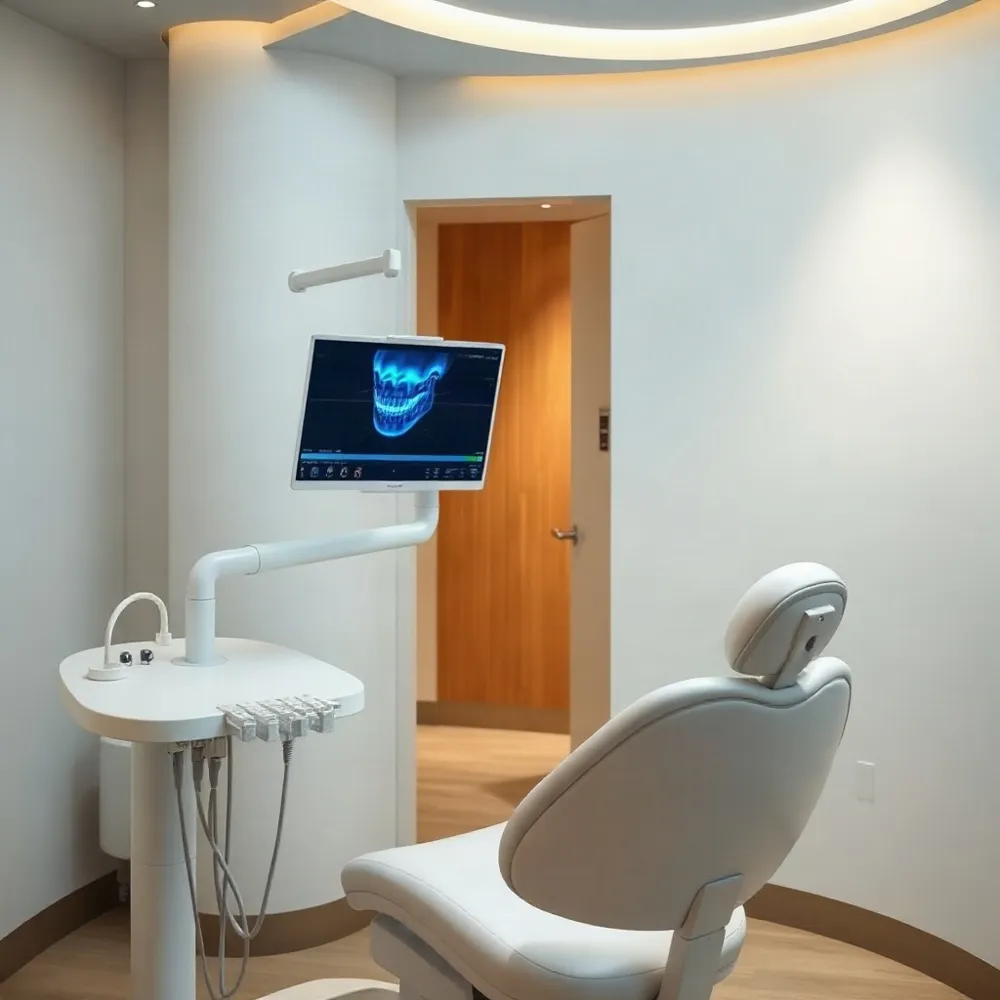 Traditional 2D dental X-rays have been the standard for decades, giving your dentist a flat, two-dimensional picture of your teeth. This is like trying to understand a three-dimensional object from just its silhouette.
A significant challenge with these flat images is that they can hide important details. Overlapping teeth, the exact position of nerves, and the true shape of your jawbone can all be obscured. Your dentist might be forced to work with an incomplete map, which can introduce uncertainty into your treatment plan.
This is where modern 3D imaging provides a complete alternative. Using advanced 3D scanning technology, a single scan captures a detailed, three-dimensional model of your teeth, roots, gums, nerves, and jawbone 3D Imaging Advancements and New Technologies in Clinical .... This advanced scan allows your dentist to rotate the image, zoom in on any area, and see how all the structures in your mouth truly relate to one another.
For the family-focused team at 3D Dental in Mastic, this means replacing old uncertainty with clear information. The detailed view provided by a 3D scan helps your dentist plan a wide range of procedures with greater accuracy and confidence, supporting gentle and effective care. This technology is especially valuable for everything from simple root canals to complex smile makeovers, ensuring that any care you receive is based on a complete understanding of your unique oral anatomy. We are committed to making this advanced technology accessible and comfortable for every member of your family, so you can feel at ease throughout your visit.
A Closer Look Inside Your Mouth
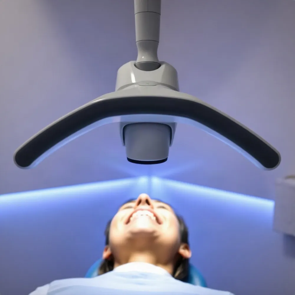 Traditional 2D X-rays give a flat picture that can hide important details. With 3D imaging, we get a complete view of your teeth, roots, jawbone, nerves, and sinuses. This modern technology helps us see things that might otherwise be missed, making your care safer and more comfortable.
3D imaging allows your dentist to see the unique structure of your mouth with great clarity. We can check the health of your jawbone and find the exact location of nerves, which is especially helpful for procedures like implants. This means we can create a treatment plan that is just right for you.
The detailed information from one scan helps us plan your care more effectively. Your dentist can preview the results of your treatment, plan the placement of crowns or veneers, and even create guides to help place implants. This reduces uncertainty and helps ensure your restoration fits well and feels comfortable.
The Power of a Single Quick Scan
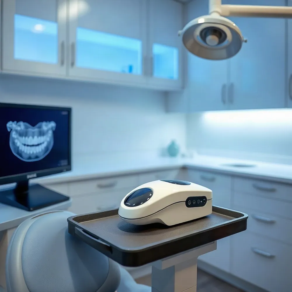 The technology that makes these precise, personalized smile plans possible is called cone beam computed tomography, or CBCT. Unlike traditional dental X-rays, which capture a single flat image, CBCT uses a cone-shaped X-ray beam that rotates around your head, capturing hundreds of images from different angles. Specialized software then assembles these into a single, highly detailed 3D model of your teeth, roots, jawbone, nerves, and sinuses. This provides your dentist with a complete view that was never possible before.
The actual scan is surprisingly quick and comfortable. You simply sit or stand in front of the machine while the scanner rotates around your head for less than a minute. There are no uncomfortable sensors to bite down on, and no messy impression materials. The entire process is non-invasive and painless, making it a much more relaxed experience than many people expect from a dental diagnostic procedure. For your safety, however, you will be asked to remove any metal jewelry, eyeglasses, or removable dental appliances before the scan, as these can interfere with the image quality.
While a CBCT scan is a powerful diagnostic tool, it is used purposefully and only when it provides clear benefits for your diagnosis and treatment plan. For routine checkups or simple fillings, a traditional X-ray is perfectly sufficient. A 3D scan is typically reserved for more complex procedures like dental implant placement, root canal therapy, orthodontic planning, or evaluating jaw issues. The practice will only recommend a CBCT scan when it meaningfully improves the accuracy of your treatment and your overall safety, ensuring you receive the right level of care without unnecessary radiation exposure.
Seeing Your New Smile Before Treatment Begins
One of the most reassuring parts of a smile makeover is knowing what the final result will look like before any work begins. 3D imaging makes this possible by giving you a clear, three-dimensional preview of your new smile. Instead of relying on verbal descriptions or flat sketches, you can see your own teeth and facial structures on a screen, from every angle, and understand exactly how the proposed changes will look and feel.
When patients can see their own anatomy in three dimensions, consultations become collaborative conversations. Your dentist can point to a specific area, explain why a particular treatment is needed, and show you how it will improve your smile. This visual clarity builds trust and helps you feel confident in the treatment plan.
At 3D Dental in Mastic, NY, Dr. Eran Arvilli uses advanced imaging technology to guide patients through every step of their smile journey. The detailed, 3D view of your teeth, gums, and jawbone ensures that nothing is left to guesswork. You and your dentist can review the plan together, making adjustments until it feels right for you.
Seeing your potential outcome before treatment removes uncertainty and replaces it with confidence. Whether you are considering veneers, crowns, or a full smile makeover, knowing what to expect helps you relax and look forward to the results. This focus on transparency and patient comfort is central to how modern digital imaging improves the dental experience.
3D imaging also makes it easier to understand the "why" behind a treatment recommendation. For example, your dentist can show you exactly where bone density is low or how a nerve runs close to the surface, turning abstract concerns into tangible information. When you understand the reasons for each step, you can make informed, confident decisions about your care.
Planning Together with Precision and Care
A smile makeover or complex restorative procedure often involves more than one dental specialty. Orthodontics, periodontics, oral surgery, and restorative dentistry each play a role, and the best results happen when these specialists work from the same detailed blueprint.
3D digital models provide that shared blueprint. Instead of each specialist relying on separate 2D X-rays and physical notes, the same high-resolution 3D image can be used by your general dentist, the oral surgeon placing an implant, and the prosthodontist designing your final restoration. This common language helps ensure every procedure is coordinated for the best possible aesthetic and functional outcome.
At 3D Dental, Dr. Eran Arvilli uses 3D imaging to bring this collaborative approach to every patient's care. By integrating orthodontic planning with restorative work and oral surgery guidance, the team can address tooth position, gum health, and bone density in one unified treatment plan. This reduces the guesswork, shortens the overall treatment timeline, and leads to more predictable, lasting results for families in the Mastic area.
Understanding the Investment in Your Smile
The cost of a 3D dental scan typically ranges from $150 to $750, depending on the area being scanned and the complexity of the case. For example, a small scan focused on a single tooth may cost between $150 and $350, while a larger scan covering the full head may run from $400 to $750.
It is wise to ask your dental team for a clear, personalized quote before any imaging is done. At 3D Dental, Dr. Arvilli and his team believe in transparent communication, so you will always know the fee in advance and can ask about bundling the scan into the overall cost of your procedure.
When it comes to insurance, coverage for 3D scans varies by plan and by the clinical reason for the scan. Scans needed for implant placement or complex root canal treatment are more likely to be covered than scans done simply for cosmetic planning. Your dental team can help check your benefits before you commit.
While the upfront cost of a 3D scan may seem like an extra expense, it is an investment in accuracy and peace of mind. The detailed view it provides reduces guesswork, lowers the chance of complications, and often shortens overall treatment time — saving you money and chair visits in the long run.
Embracing Modern Technology with a Warm Touch
From a single, quick scan to a virtual preview of your new smile, 3D imaging transforms the dental experience. It brings greater accuracy, comfort, and peace of mind to every step of your treatment.
At 3D Dental, Dr. Eran Arvilli and the team combine this advanced technology with the gentle, personal care that makes our practice a trusted partner for families across Suffolk County. We take the time to walk you through every image and every option, answering all your questions in a calm, judgment-free setting.
Whether you are visiting us for a routine checkup or a complete smile makeover, you can feel at ease knowing that modern diagnostics and a warm bedside manner go hand in hand. Our bilingual team welcomes Spanish-speaking patients, ensuring everyone feels understood and respected.
Your smile is unique, and your care should be too. We invite you to ask questions, share your goals, and discover how gentle, precise dentistry can make a lasting difference in your life.